
Go DeepSmall animal deep image with non-invasive measurement in visible region
|
|
小動物腫瘤斷層影像開發
|
Small animal multimodality 3D imaging system: fluorescence diffuse optical tomography, bioluminescence tomography, and ultrasound image for in vivo small animal study.
Fluorescence diffuse optical tomography (FDOT) is a non-invasive measurement that uses the surface light distribution to reconstruct the FDOT in vivo and has been used widely for small animal studies. In this topic, we developed a small animal multimodality three-dimensional (3D) imaging system combining FDOT, bioluminescence tomography (BLT), and ultrasound (US) imaging. The advantages of using this system are a non-invasive, easy-to-use, and good contrast to soft tissue. We validated the system with porcine tissue and nude mice. The reconstruction results show the FDOT-US system can effectively localize the tumor and drug metabolism. It is very helpful for the study of tumor location and the development of cancer drugs in small animal studies. 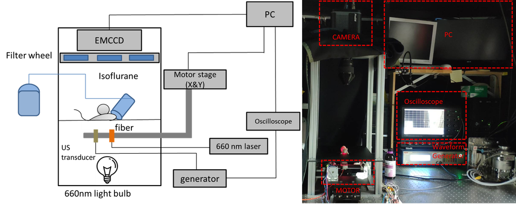
Schematic of the FDOT-US system 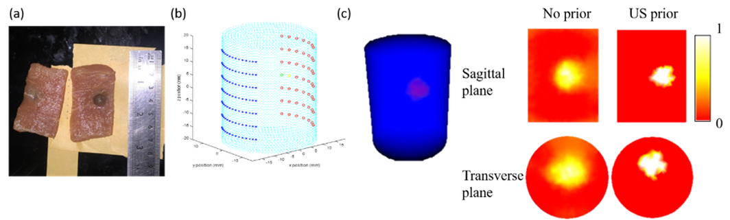
FDOT Reconstruction of porcine tissue a. A cylinder shape porcine tissue with a fluorophore gel ball. b. The arrangement of the light sources and detectors. c. FDOT reconstruction with US prior. 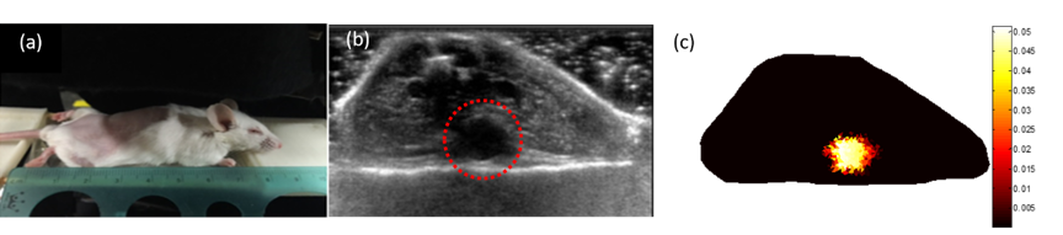
FDOT Reconstruction of small animal a. A nude mice Implanted with fluorescent tube. b. The US image of the fluorescent tube (red circle) containing section. c. FDOT reconstruction with US prior. 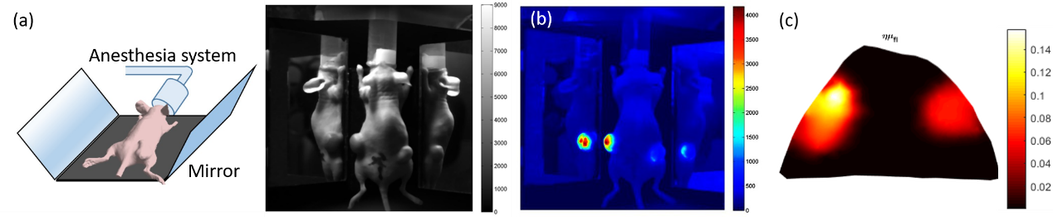
BLT Reconstruction of small animal a. Experimental set-up of BLT system b. Bio-luminescence planar image used to locate the tumor in the mouse c. BLT reconstruction with US prior |
